Early Stage Ovarian Cancer Color Normal Ovary Doppler Ultrasound

Selection Of Hens With Normal Ovaries Or Ovarian Tumors Using A C Download Scientific Diagram

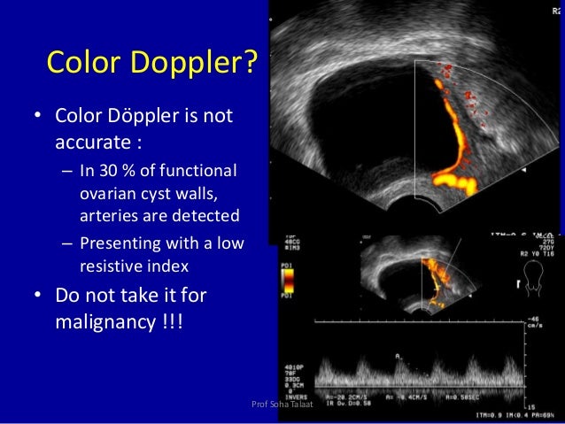
Ovarian Cancer Color Doppler

Ultrasonographic Color Doppler Appearance Of The Ovaries With Moderate Download Scientific Diagram

Color Doppler Image Of The Right Ovary Demonstrates The Typical Ring Download Scientific Diagram
A Gallery Of High Resolution Ultrasound Color Doppler 3d Images Ovaries
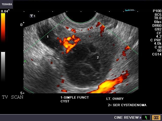
A Gallery Of High Resolution Ultrasound Color Doppler 3d Images Ovarian Masses
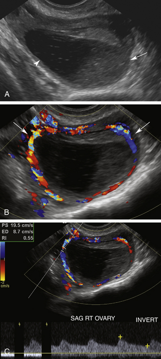
Using the power of 3d ultrasounds doppler evaluation better illuminates the presence of rich vascularity color score 3 4 which is predominant in metastatic cancers as well as the central localization of vessels in a tumor which are important parameters that contribute to the differentiation between benign and malignant ovarian masses.
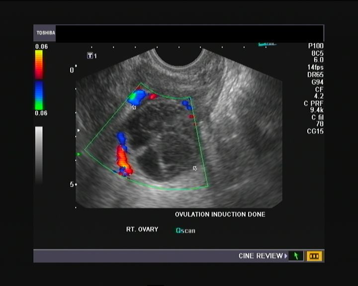
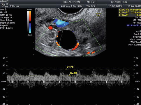
Early stage ovarian cancer color normal ovary doppler ultrasound.
Duplex Ultrasound Evaluation Of The Uterus And Ovaries Radiology Key
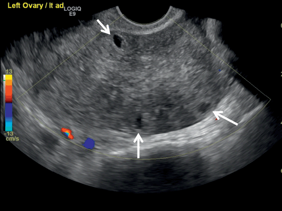
The Normal Ovary Changes In The Menstrual Cycle Radiology Key

Detection Of Ovarian Tumors In Chicken By Sonography Barua 2007 Journal Of Ultrasound In Medicine Wiley Online Library
Doppler In Gyneacology Dr Muhammad Bin Zulfiqar
Source : pinterest.com
